
A prototype from Siemens Healthineers has shown in clinical trials the potential to increase spatial resolution, while reducing X-ray exposure to patients, improving image quality by lowering image noise and artifacts, and distinguishing multiple contrast agents.
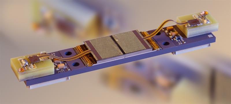
Siemens Healthineers had asked CEA-Leti to design, integrate, manufacture, and test a new generation of PCDM that will become mature enough to be integrated into an X-ray CT scanner prototype.
X-ray PCDMs based on cadmium telluride (CdTe) allow simultaneous acquisition of high-spatial-resolution and multi-energy images. Higher spatial resolution improves image quality by using a small-pixel size detector, while image noise can obscure features, and artifacts can mimic them. Multi-energy provides colour images, compared to grey-level images of conventional detectors, and allows a precise determination of the atomic number of any chemical elements present in the body.
“The successful collaboration with CEA-Leti allowed Siemens Healthineers to prototype what the med- tech company sees as the future of detector modules for whole-body CT,” explained Jean-Michel Casagrande, the project manager for medical X-ray imaging at CEA-Leti.
X-ray CT scanners use computer-processed combinations of many X-ray measurements taken from different angles to produce cross-sectional images of scanned objects. Current X-ray CT scanners produce images with energy-integrating detectors (EIDs), which are based on indirect conversion technology: X-ray photons are first converted into visible light using scintillator material, then visible photons produce electronic signals using a photodiode. PCDMs, on the other hand, directly convert X-ray photons into electronic signals with a higher conversion yield.
In addition, EIDs register the total energy deposited in a pixel during a fixed period of time without discriminating between low- and high-energy photons. This produces a monochrome X-ray image that shows the density of the body’s organs. PCDMs count each photon, which improves the contrast-to-noise ratio of the image, and the energy classification of the detected photons can then be used to produce a colour image that allows a precise determination of the atomic number of any chemical elements and a distinction of multiple contrast agents present in the body.
Finally, the detector module’s very high spatial resolution generates clearer images of very-fine structures, such as small airways in the lungs, trabeculae in bones, and thin wires in coronary stents than current scanner technology.
“The idea of Siemens Healthineers to integrate PCDMs in the future generation of X-ray CT scanners was new and no available technology existed when CEA-Leti began working on this,” said Loick Verger, the industrial-partnership manager for X-ray imaging at CEA-Leti. “The technical challenge – low noise at a very high counting rate, two energy classifications, and sufficient maturity to be integrated in an X-ray CT scanner – was tremendous.”
Verger explained that CEA-Leti used its simulation tools to design the geometry of the detector, chose a semiconductor based on CdTe, designed the electronic readout circuit, and then was able to propose a reliable CdTe electronic assembly technology.
“Researchers at the Mayo Clinic in the U.S. have evaluated Siemens Healthineers’ photon-counting detector system’s performance in phantoms, cadavers, animals, and humans. Images of more than 300 patients produced with this technology consistently demonstrated that the theoretical benefits of this type of detector technology yield a number of important clinical benefits,” said Cynthia McCollough, professor of Medical Physics and Biomedical Engineering at the Mayo Clinic.
“Publications by our research team have shown improved spatial resolution, decreased radiation or iodine contrast dose requirements, and decreased levels of image noise and artifacts,” McCollough said. “Additionally, the ability to simultaneously acquire multiple 150-micron-resolution datasets, each representing a different energy spectrum, is anticipated to lead to new clinical applications.”


