Regular eye checks are important because sight loss is avoidable in 50% of cases; people might not be aware they have eye problems until it starts affecting their vision. Catching problems early and getting treatment is key to preventing sight loss.
Eye testing, at least in my experience, has been a low tech activity. You wear a frame that could double as a device of torture into which the optician fits a combination of lenses that not only corrects your vision, but also determines whether you suffer from astigmatism, amongst other things.
The traditional eye test is based around the Snellen chart. Developed in 1862 by Dutch ophthalmologist Herman Snellen, the chart displays 11 lines of block letters. One very large letter at the top of the chart is acccompanied by rows of diminishing sizes. It’s where the phrase ‘20/20 vision’ comes from. The chart should be read from a distance of 20ft; if you can read the line designated 20/20 – said to be the smallest line that a person with normal visual acuity can read at that distance – then you have ‘20/20 vision’.
In case you’ve been wondering, BS 4274-1:2003 stipulates that only the letters C, D, E, F, H, K, N, P, R, U, V and Z should be used for the testing of vision, based upon equal legibility of the letters.
But my latest visit to the optician showed technology is becoming more important, not only in determining the prescription and the internal condition of the eye, but also in the fitting of the frames.
Benedikt Wurm is a product manager with Heidelberg Engineering, a high tech medical device company which designs and manufactures diagnostic instruments for use by eye care professionals around the world. He said: “Examining the inside of the eye probably started a couple of hundred years ago, when doctors started to look at the retina, cornea and iris. At that time, they tried to look through the pupil using a lens to see what was going on.”
And there is a lot going on inside the eye, Wurm noted. “You will find a unique pattern within the eye because nerve fibres coming from the brain are not covered by anything; it’s one of the few organs in the human body where you can see things happening.”
More recently, the inside of the eye was explored in greater details using cameras. “The patient was examined, a photo taken, followed by another when they returned. The two were then compared to see whether there were any changes.”
While that approach provided some information, Wurm said cameras were a problem. “The patient’s pupil can be small and may not widen in certain conditions, even with the use of eyedrops.”
A big step forward came with the use of laser scanning, rather than photography. “It’s possible to use several lasers to get a better image quality,” he said. “A confocal laser system allows professionals to get a sharp image of the back of the eye.”
The use of different lasers is important as this allows a range of information to be gathered. “For example,” Wurm noted, “infra red laser light penetrates beneath the surface.”
Heidelberg’s Spectralis features five lasers: blue (486nm); green (518nm); near IR (786nm); far IR (815nm); and superluminous (870nm). It combines optical coherence tomography (OCT) with confocal scanning laser ophthalmoscopy to provide new ways to image the structure and function of the eye.
OCT is an imaging technology, similar to ultrasound and MRI, in which reflected light is used to produce detailed cross sectional and 3D images of the eye. “You get a 3D image similar to a CT scan,” Wurm explained. “It’s the biggest thing that’s happened in opthalmology for some time.”
An early version of this technology, time domain OCT, used a moving reference mirror to measure the time it took for light to be reflected. This relatively slow, mechanical process limited the amount of data that could be captured, as well as image quality.
Wurm said: “Time domain OCT had poor resolution, motion artefacts and was complicated to use. Once the mirror is replaced with a broad spectrum light source, better quality images can be acquired more quickly.
Early laser based instruments allowed 30k scan/s to be captured, which has since improved to 85k scan/s. “Spread spectrum technology increases this to more than 100k scan/s,” Wurm said.
A further improvement on this approach is swept source OCT, which Wurm says is not available commercially as yet.
Like the cameras in the early days, the laser based instruments image the retina through the pupil. “Opthalmologists can see the choroid – the vascular layer of the eye beneath the retina – and examine the vitreous humour,” Wurm said. “In fact, OCT is the ‘gold standard’ for detecting vitreous detachment, which can cause retinal detachment.”
Asked why scan speed was so important, Wurm said it was because the eye moves and because patients don’t want to stare into instruments.“Even if you’re looking at one point, the eye is still moving,” he said. “If the object is moving faster than you can image it, there will be motion artefacts and opthalmologists could ‘see’ something that isn’t there. With the wrong information, it’s possible to make the wrong diagnosis.”
The retina is scanned in 24ms ‘bursts’ and this is faster than most involuntary eye movements; so called saccades – French for jerks – have a period from 30 to 50ms, while voluntary eye movements take more than 150ms.
A standard lens captures an image of a 9 x 9mm area of the retina, while optional lenses expand the field of view.
Wurm said there is little post processing involved. “For a superficial image, the back of the eye is scanned on a point by point basis and what comes out is registered.”
That image has a resolution of 1536 x 1536 pixels. “This is normalised using a Fourier transformation,” Wurm continued, “but there is no further manipulation of an OCT image because it’s based on interferometry.”
In terms of vertical resolution, the system will produce a 496 pixel deep cross section. “That doesn’t sound a lot,” Wurm said, “but that gives professionals all the information they need. Each pixel equates to a depth of about 4µm, so the different layers can be resolved.”
Although specialised, devices such as Spectralis are becoming more widely used. “It’s a ‘must have’ for specialists and clinics,” Wurm said. “In the UK, optometrists are opening up to the technology – traditionally, they haven’t adopted new technology very quickly. Price is decreasing over time, driven by competition, but also because technology is less expensive,” he concluded.
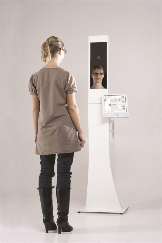
More accurate measurements According to equipment and lens specialist Essilor, every eye is not only structurally different, but also moves differently. Using its VisiOffice system, a unique ‘eyecode’ can be created, with 20 times more measurements taken in 30s than can be done in a standard eye test. Visioffice, said to provide results free of parallax, can measure to within 0.1mm for distance measurements and to the nearest degree for angles. Amongst the measurements taken are the eye rotation centre and the distance between the front of the eye and the back of the lens; something which couldn’t be done before. |
Imaging technology developed at the University of Illinois could help eye doctors to see individual cells in the back of a patient’s eye, thanks to imaging technology. Such detailed pictures of the cells, blood vessels and nerves could enable earlier diagnosis and better treatment for degenerative eye and neurological diseases. The approach uses computational techniques to all the rods and cones – cells in the back of the eye that detect light – to be seen more clearly. Professor Stephen Boppart said: “The eye has always been a bit of a challenge to image; it’s a very complicated organ. There are many microscopic structures that are hard to see. Many diseases that affect vision also start at the microscopic level, so being able to see those early changes is going to lead to better, earlier treatment.” The prevailing imaging technique in ophthalmology – optical coherence tomography (OCT) – is useful for general imaging of the eye, but cannot focus down to the scale of individual rods and cones. In addition, OCT images are often blurred by the eye’s imperfections and motion. The Illinois team has developed a computational adaptive optic approach in which complex algorithms are applied to OCT data, yielding high resolution, real time images. The team is initially focusing on using computational adaptive optics to track age related macular degeneration and multiple sclerosis. “I think computational adaptive optics can be really helpful to the clinical community,” said Fredrick South, one of the researchers. “It could give ophthalmologists information that, currently, they have to infer from other measurements. They can’t directly look at the photoreceptors and watch them die off during macular degeneration, for example – they just have to guess what’s going on. “It could be possible to use computational adaptive optics in these real world applications both for diagnosis of disease and tracking of treatments.” |
 Computational approach reveals rods and cones
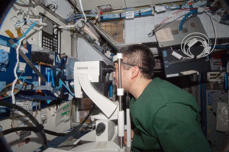
Being in space changes your eyes Following research that pointed to a permanent vision risk for astronauts living without gravity for at least six months, astronauts on the International Space Station (ISS) are studying why weightlessness alters vision. “After a few weeks aboard the [ISS],” said Bob Thirsk, who spent six months on the ISS in 2007, “I noticed that my visual acuity had changed. My distant vision was not too bad, but I found that it was more difficult to read procedures. I also had trouble focusing cameras manually.” Amongst the problems discovered are a flattening of the globe, swelling of the optic nerve and choroidal folds. “Swelling of the optic nerve is a serious problem; if it’s chronic, that could cause some permanent damage to the eyes,” said Dr Robert Gibson, who conducted the survey. “We do see some change to the anatomy, to the structure of the eye; we see little wrinkles to the retina and the back of the eye, and change in the shape of the eyeball that may be permanent for some.” Heidelberg Engineering’s Benedikt Wurm said NASA now wants to monitor what’s happening in the eye. “So modified systems have been sent into space that allow astronauts to be examined during their missions.” Examinations before and after missions allow all changes to be discovered. Wurm said two systems were transported to the ISS on the ATV4 supply vessel; one of which is a back up. “They have to launch proof,” he noted, “but are being used for examinations on the ISS on a regular basis.” |