
Their new technique relies on optical fibre technology to provide new sensors for photoacoustic imaging. It uses fibre-optic ultrasound detection, exploiting the acoustic effects on laser pulses via the thermoelastic effect - temperature changes that occur as a result of the elastic strain.
"Conventional fibre optic sensors detect extremely weak signals by taking advantage of their high sensitivity via phase measurement," said Long Jin from the Institute of Photonics Technology at Jinan University.
These same sorts of sensors are used in military applications to detect low-frequency (kilohertz) acoustic waves. But it turns out that they don't work so well for ultrasound waves at the megahertz frequencies used for medical purposes because ultrasound waves typically propagate as spherical waves and have a very limited interaction length with optical fibres. The new sensors were specifically developed for medical imaging, Jin explained, and can provide better sensitivity than the piezoelectric transducers in use today.
The group designed a special ultrasound sensor that's essentially a compact laser built within the 8-micron-diameter core of a single-mode optical fibre. "It has a typical length of only 8 millimetres," Jin said. "To build up the laser, two highly reflective grating mirrors are UV-written into the fibre core to provide optical feedback." This fibre then gets doped with ytterbium and erbium to provide sufficient optical gain at 1,530nm. They use a 980nm semiconductor laser as the pump laser.
"Such fibre lasers with a kilohertz-order linewidth - the width of the optical spectrum - can be exploited as sensors because they offer a high signal-to-noise ratio," said Assistant Professor Yizhi Liang of Jinan University.
The ultrasound detection benefits from the combined technique because side-incident ultrasound waves deform the fibre, modulating the lasing frequency. "By detecting the frequency shift, we can reconstruct the acoustic waveform," Assist Prof. Liang said.
The team does not demodulate the ultrasound signal, extracting the original information, using conventional interferometry-based methods or any additive frequency locking. Rather, they use another method, called "self-heterodyning," where the result of mixing two frequencies is detected. Here, they measure the radio frequency-domain beat note given by two orthogonal polarization modes of the fibrecavity. This demodulation also intrinsically guarantees a stable signal output.
The researchers used a focused 532nm nanosecond pulse laser to illuminate a sample and excite ultrasound signals. By placing a sensor in a stationary position near the biological sample, they said they were able to detect optically induced ultrasound waves.
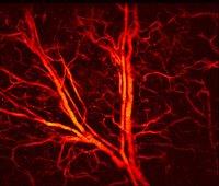
"By raster scanning the laser spot, we can obtain a photoacoustic image of the vessels and capillaries of a mouse's ear," Jin said. "This method can also be used to structurally image other tissues and functionally image oxygen distribution by using other excitation wavelengths -- which takes advantage of the characteristic absorption spectra of different target tissues.
"The development of our laser sensor is very encouraging because of its potential for endoscopes and wearable applications," he continued. "But current commercial endoscopic products are typically millimetres in dimension, which can cause pain, and they don't work well within hollow organs with limited space."
| A photoacoustic microscope image of blood vessels and capillaries in a mouse’s ear (size: 2.7x2.7mm2). Credit: Long Jin |


